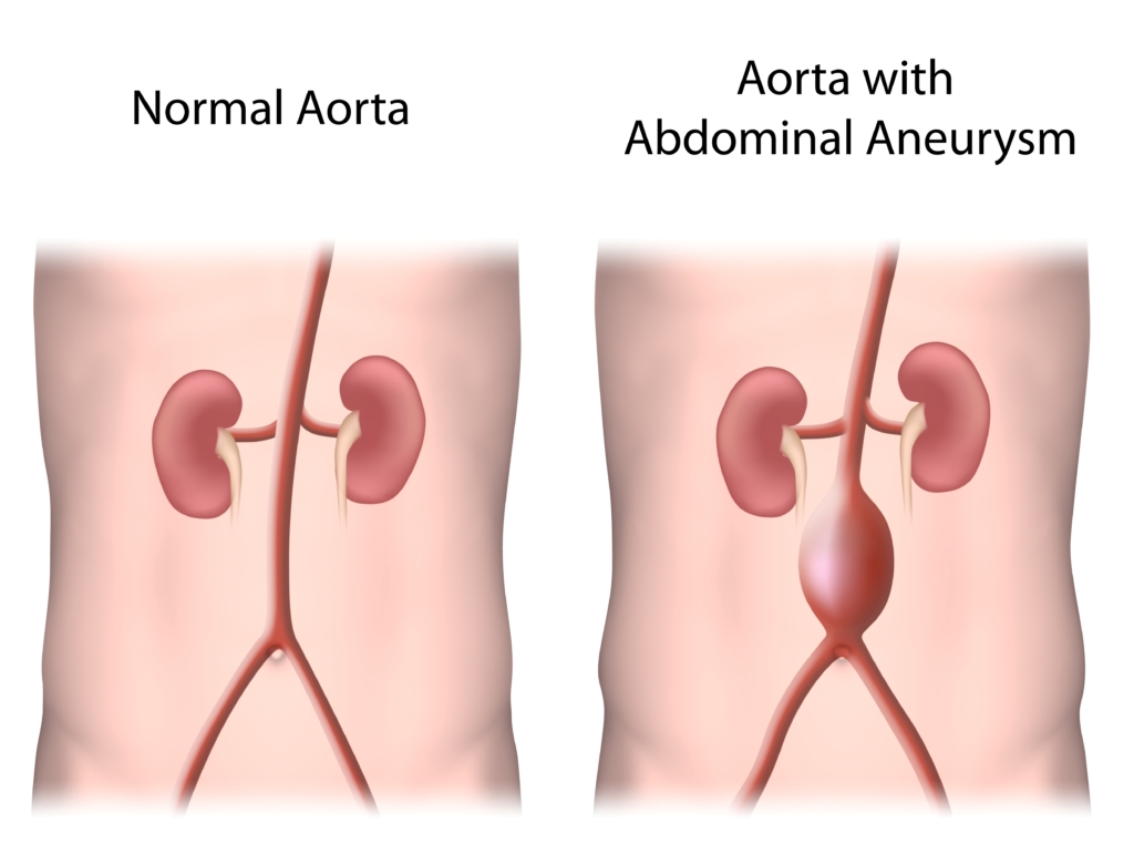
Aortic aneurysms are dilatations (or swellings) of the aorta, which is the main artery from the heart supplying blood to the body. They most commonly occur in the abdomen although they can occur in the chest. They do not cause symptoms usually until they rupture, which is often fatal. They are diagnosed by performing an abdominal ultrasound or CT scan. If there is an aortic aneurysm, then there is a possibility that other arteries can also have an aneurysm, most commonly involving the leg artery behind the knee.
The risk of rupture is directly related to the maximum diameter, and treatment is advised when the aneurysm reaches 5.5cm, as this is the size where the risk of rupture overtakes the risk of treatment. The natural history of aortic aneurysms is to slowly enlarge (about 3-5mm per year) although they can remain static for years. Once an aneurysm is diagnosed, then 6-monthly or annual scans should be performed to monitor expansion rate. This can be done in our vascular ultrasound lab.
Symptoms: AAAs often grow slowly and very often there are no symptoms. An aneurysm may start small and stay small or expand quickly. Predicting how fast it may grow is difficult. Once diagnosed the patient will be put on a vascular ultrasound surveillance schedule. As an AAA enlarges some patients may notice the following
- A pulsating feeling near the navel
- A constant pain in the abdomen
- Back pain


